The Keys to Cheese: Does This Cheese Melt?

Melted Cheese [Photo Credit: Pittaya Sroilong]

Melted Cheese Frize [Photo Credit: Pittaya Sroilong]

Whether you are making cheese fries, grilled cheese sandwiches, quesadillas, baked cheese bites, or homemade mac and cheese, choosing the right type of cheese can make or break these comfort foods. The key to all of these dishes is cheese that produces an even and homogenous melt. Cheeses like Cheddar, Mozzarella, and Gruyere are used often. If you aren’t feeling adventurous, you could just memorize the names of these greatest hits. However, if you want to experiment and change the melty cheese game, you’re going to have to understand why these cheeses work.

Let’s first examine what happens to cheese as it melts. The interactions of casein (milk proteins) and calcium help define its solid structure. When solid, caseins are bound together in large branching porous protein networks that entrap milkfat and water. Calcium (as calcium phosphate) acts as a bridge to stabilize these networks. When you apply heat to a cheese, melting occurs in two stages. First, at around 90 ˚F, milkfat is released1. This is because hydrophobic (water-repulsive) interactions between casein molecules increase under heat2. These interactions force out water molecules and the space between casein molecules increases allowing milkfat, which melts at this temperature, to escape. If you’ve put cheese on a burger that’s being grilled, you may see little sweat beads of liquid form on the cheese in the early stages of melting. The second stage happens at about 40 to 90 degrees higher, at around 130 – 180˚F3. At this point, the casein proteins do not break down, but rather, the increased movement of the proteins, resulting from the heat, allows for the proteins to act more fluid-like and the cheese melts.

There are many factors that control melting and explain why melting temperatures vary by as much as 50 degrees. No one factor defines a cheese’s melting properties as these factors can interact.

Moisture and Fat

Cheese with higher moisture and fat content tends to have lower melting points. For example, high moisture cheeses like Mozzarella melt around 130 ˚F and low moisture cheeses like Swiss melt at 150 ˚F 2. First, as previously highlighted, the milkfat and water portion of the cheese react to heat at lower temperatures than the proteins. Accordingly, with more moisture and fat present in a cheese, greater proportions of the cheese are susceptible to melting at lower temperatures. When the fat becomes liquid, it can no long provide support for the protein networks. Secondly, increased moisture and fat means that the casein proteins are more spread out and the mesh size (gap between proteins) is larger. This means there are fewer connections (bound calcium bridges) between proteins networks making melting more likely to occur at lower temperatures.

You may not know the exact moisture and fat content of every cheese variety without looking at a label, but intuitively, softer cheeses have more moisture and fat. Additionally, younger cheeses generally have more moisture so they also tend to melt more uniformly and evenly.

Acid Content

Chesses typically melt homogenously and evenly around a pH of 5.0 – 5.44. This is related to the calcium bridges. At too high a pH (pH > 6), too much calcium is present as bound calcium phosphate and the protein is too tightly bound to melt. With lowered pH, the calcium phosphate bound to the casein is replaced by hydrogen (H+), allowing for more movement among proteins.2 At around a pH of 5.0 – 5.4, there is a sufficient number of calcium present as bridges to allow for melting. At too low a pH (pH < 4.6), too many calcium bridges are lost and proteins aggregate and are unable to flow and melt evenly.

Lastly as a caveat, the factors being highlighted are specific to rennet-set cheeses, and not acid-set cheeses. Acid-set cheeses like queso fresco, paneer, and ricotta are not generally used, as they don’t produce even melts4. This results from the way they were made. In cheese making, you have two options for separating the solid curds (primarily casein proteins) and the liquid whey; Use rennet (an enzyme derived from the intestines or baby goats and cows) or use an acid (like vinegar or lemon juice).

When they are free floating in liquid milk, casein proteins have a slightly different molecular structure than when they are in cheese. In milk, caseins stick together in small clusters (micelles) that have negative charges on their surface. Since negative charges repel each other, these micelles won’t combine. Adding acid to heated milk lowers the pH, which neutralizes the negative charges on the micelles; therefore the casein micelles can aggregate. In contrast, using rennet to set cheese is a more targeted approach. In this process, an enzyme contained in rennet called chymosin, selectively removes negatively charged portions of the casein micelles and allows the micelles to clump.

In an acid-set cheese, calcium bridges are never formed as a result of the acidic environment used to generate the cheese5. These cheeses are only held together in protein aggregates rather than protein networks with calcium bridges and don’t produce the even melt desired.

Bottom Line:

Rennet-set cheeses with high moisture and fat are the best cheeses for melting as they melt evenly and consistently.

But don’t fret if you still want to harness the flavor of other cheeses (especially older or drier cheeses)! You have options: Try using a cheese blend with a higher proportion of the better melting cheeses and a small proportion of the other cheeses. For example, this recipe uses a 1:4 ratio. Experiment! You now know the keys for melty cheese!

References cited

  1. Schloss, Andrew and David Joachim. “The Science of Melting Cheese” http://www.finecooking.com/item/64019/the-science-of-melting-cheese
  2. Johnson, Mark. “The Melt and Stretch of Cheese” https://www.cdr.wisc.edu/sites/default/files/pipelines/2000/pipeline_2000_vol12_01.pdf
  3. Mcgee, Harold. On Food and Cooking. 2004 “Cheese” (57 – 67).
  4. Tunick, Michael. The Science of Cheese. 2013 “Stretched Curd Cheeses, Alcohols, and Melting” (82 – 91).
  5. Sargento Food Service. “Cheese Melt Meter” http://www.sargentofoodservice.com/trends-innovation/cheese-melt-meter/
  6. Achitoff-Grey, Niki. “The Science of Melting Cheese” http://www.seriouseats.com/2015/08/the-science-of-melting-cheese.html

Vince ReyesAbout the author: Vince Reyes earned his Ph.D. in Environmental Engineering at UCLA. Vince loves to explore the deliciousness of all things edible.

Read more by Vince Reyes


Structural Changes in Chocolate Blooming

Is there anything more disappointing than finding a chocolate bar in the back of the desk drawer, anticipating a tasty treat, then unwrapping the bar only to find a dull, grey haze has overtaken your dear candy? Seeing as bloomed chocolate is still edible, yes, there are many things more disappointing than that. But surely you’re curious about how chocolate that was once shiny and perfect came to be filmy and rough. Chocolate blooming, the process that produces the white-grey film that appears on the surface of an old chocolate, is due to molecular migration. More specifically, this imperfection is caused by the movement of fats to the surface of the chocolate followed by a subsequent recrystallization. In a paper published by Applied Materials & Interfaces, a team of researchers dedicated to keeping our chocolates blemish-free has clarified the precise mechanisms that cause chocolate blooming.

The main fat in chocolate is cocoa butter, which is solid at room temperature and melts at 37 degrees Celsius. The proportion of solid to liquid cocoa butter depends on the lipid composition, which depends on which specific triglycerides are present. The solid to liquid proportion also varies with the storage conditions of the chocolate.

As proposed by Aguilera et al, scientists who study this chocolate blooming, consider chocolate as a particulate medium of fat-coated particles such as cocoa solids, sucrose, and milk powder, all suspended in a fat phase with the aid of an emulsifier, which helps to mix fats and oils with water, which usually repel each other. There are six crystallographic polymorphs of cocoa butter molecules, that is, there are six ways the molecules can organize themselves. The structural stability of these polymorphs increases from 1- 6; form 1 is the best at forming solid butter at room temperature, while form 6 tends to arrange in the loose bonds of a liquid. Form 5 is the main form in chocolate, as it possesses the most aesthetically desirable properties. While the phenomenon of blooming is well known to result from melting and recrystallization of chocolate into a less desirable polymorph, it has been unclear how fat moves through the chocolate particle network: Does it move along the fat-particle interface? Does it diffuse through the fat phase (cocoa butter), or through the matrix of assorted particles?

Possible lipid migration pathways in chocolate - Reinke et al

Possible lipid migration pathways in chocolate – Reinke et al

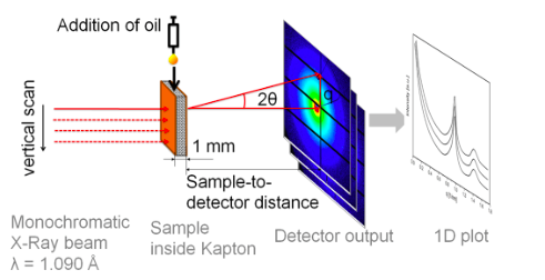
In this experiment, researchers used synchrotron microfocus small-angle X-ray scattering to determine the preferential migration pathway of the cocoa butter molecules surrounded by three different soild components (cocoa solids, skim milk, and sucrose). This technique allows researchers to record the scattering of x-rays through a sample with defects in the nanometer range. They can then extrapolate information about the material’s macromolecules, their shapes and sizes up to 125 nanometers, and distances between partially ordered materials, such as pore sizes. For this experiment, this method is better than more traditional macroscopic techniques as the sample does not need to be dissected in order to examine it, therefore the same sample can be continually analyzed.

Sketch of the experimental setup - Reink et al

Sketch of the experimental setup – Reink et al

The researchers prepared and tempered four different chocolate samples. An initial scattering of x-rays and data collection was performed before the addition of sunflower oil, then 10 uL of oil was pipetted onto the chocolate surface, and a second scan was performed. Images of the droplet were captured through a high-speed camera. These scans were repeated at 5, 10, and 30 minutes after oil addition, and again after 1, 2, 5, and 24 hours.

The results obtained suggest that oil is migrating through pores and cracks in the solid structure driven by capillarity within seconds. This means that the oil can flow in narrow spaces in opposition to gravity. Then chemical migration through the fat phase occurs. The oil doesn’t traverse the fat-particle interface, nor does it move through the matrix of solid particles. This migration disrupts the crystalline cocoa butter, which induces softening.

Because the most immediate migration of oils occurs through the material porous structure, the formation of chocolate bloom could be prevented by minimizing pores and defects in the chocolate matrix. To prevent the longer-term effects of chemical migration of lipids, one must minimize the content of non-crystallized liquid cocoa butter. Tempering chocolate lends to crystalline structures that resist migration, as will reducing the liquid fat content. However, to ensure that you never encounter a sad hazy chocolate again, we recommend eating all chocolate goods expeditiously.

Works Cited

  1. Tracking Structural Changes in Lipid-based Multicomponent Food Materials due to Oil Migration by Microfocus Small-Angle X-ray Scattering. Svenja K. Reinke, Stephan V. Roth, Gonzalo Santoro, Josélio Vieira, Stefan Heinrich, and Stefan Palzer. ACS Applied Materials & Interfaces 2015 7 (18), 9929-9936. DOI:10.1021/acsami.5b02092
  2. Aguilera, J. M.; Michel, M.; Mayor, G.Fat Migration in Chocolate: Diffusion or Capillary Flow in a Particulate Solid?—A Hypothesis PaperJ. Food Sci. 2004, 69, 167–174


Elsbeth SitesAbout the author: Elsbeth Sites received her B.S. in Biology at UCLA. Her addiction to the Food Network has developed into a love of learning about the science behind food.

Read more by Elsbeth Sites


Caffeine vs. Chocolate: A Mighty Methyl Group

Guest post by Christina Jayson

Photo credit: Lisa Townley (left); Pyogenes Gruffer (right), Flickr.

Photo credit: Lisa Townley (left); Pyogenes Gruffer (right), Flickr.

When my organic chemistry professor told me that the main molecular component of chocolate, theobromine, differs from caffeine only by the absence of one methyl group I was delighted: I could skip an entire step in caffeine metabolism, avoid the bitter taste of coffee, and increase my chocolate consumption. It seemed to make sense that as the caffeine I drank was metabolized by removing the methyl group, caffeine would convert to theobromine (the main compound of chocolate) (Figure 1). At the molecular level, a methyl group is a carbon with three hydrogens attached. It may seem simple, but a methyl group is an integral part of chemistry, biology, and biochemistry. For example, additional methyl groups can help a molecule to cross the blood-brain barrier and enter our brain – this barrier protects our brain from foreign molecules traveling in the blood that can be harmful [1, 2]. In the case of caffeine, it turns out that the extra methyl group on the molecule is what makes coffee active on our central nervous systems and an “energy stimulator,” while chocolate functions as a sweet treat and smooth muscle stimulator.

Figure 1: During the metabolism of caffeine in the body, the methyl group (highlighted by the yellow box) is removed from caffeine and it is converted to theobromine (Modified from Wolf LK, 2013) [9].

Figure 1: During the metabolism of caffeine in the body, the methyl group (highlighted by the yellow box) is removed from caffeine and it is converted to theobromine (Modified from Wolf LK, 2013) [9].

So how do these two molecules act on different parts of the body, making coffee the substance of choice over chocolate bars when midterm season hits?

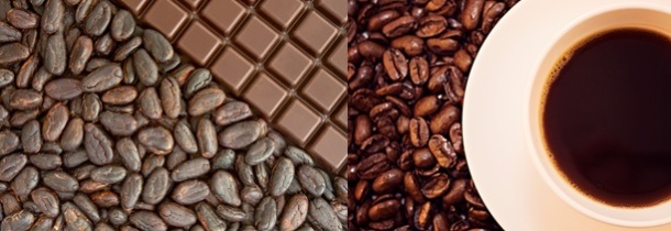
Caffeine is mostly derived from Coffea Arabica, or coffee beans, and seeds [3]. It is predominantly a central nervous stimulant, though it also stimulates cardiac and skeletal muscles and relaxes smooth muscles. Chocolate, or theobromine, is found in products of Theobroma cacao, or cocoa plant seeds (Figure 2). Much like caffeine, theobromine is a diuretic; however it mainly acts as a smooth muscle relaxant and cardiac stimulant [3]. While these two compounds have similar effects, the key difference is that caffeine has an effect on the central nervous system and theobromine most significantly affects smooth muscle [4]. In behavioral studies, caffeine intake improves self-reported alertness and mood over a period of 24 hours [5]. Theobromine produces mild positive effects in pleasure, but does not affect attention or alertness in moderate doses compared to caffeine [6].

Figure 2: Chocolate (left) is made from Theobroma cacao, or cacao plant seeds and contains theobromine (PC: Nic Charalambous). Coffee (right) is made from Coffea Arabica, or coffee beans, and seeds and contains caffeine (Photo credit: JIhopgood/Flickr).

Figure 2: Chocolate (left) is made from Theobroma cacao, or cacao plant seeds and contains theobromine (PC: Nic Charalambous). Coffee (right) is made from Coffea Arabica, or coffee beans, and seeds and contains caffeine (Photo credit: JIhopgood/Flickr).

But the true difference in the compounds lies at the molecular level. Both caffeine and theobromine belong to the methylxanthine chemical family. These chemicals act as stimulants of the nervous system, most notably by binding to adenosine receptors in the brain and thereby blocking adenosine from binding to the receptors [7]. Adenosine binding to adenosine receptors normally reduces neural activity, so the antagonistic action of caffeine and theobromine prevents this activity reduction (Figure 3). The increased energy and alertness that we connect to massive coffee consumption is due to the caffeine preventing your body from responding to signals that tell it to slow down or de-stimulate. Ever felt your hands jitter uncontrollably after too many shots of espresso?

Figure 3: Caffeine molecules (C) compete with adenosine molecules (A) to bind to the adenosine receptors in the brain (Schardt, 2012) [10].

Figure 3: Caffeine molecules (C) compete with adenosine molecules (A) to bind to the adenosine receptors in the brain (Schardt, 2012) [10].

Experiments show the activity of caffeine on the nervous system is stronger than theobromine [7]. Caffeine and theobromine compete with adenosine to bind to the same adenosine receptor. Studies have shown that caffeine molecules are better able to compete with adenosine to bind adenosine receptors than theobromine – caffeine binds these receptors with two to three times higher affinity than theobromine [8].

To gain access to the different locations of the adenosine receptors throughout the body, the extra methyl group on caffeine ends up coming in handy. Because caffeine has three methyl groups instead of two like theobromine, it more easily crosses the blood-brain barrier. In crossing the blood-brain barrier, caffeine can act on the central nervous system. So while theobromine can act as a heart stimulant and smooth muscle relaxant, caffeine – boasting its extra methyl group – has access to the neurons of the central nervous system and can consequently enhance physical performance and increase alertness.

Photo credit: Chris Swift, Rogers Family Co [11]

Photo credit: Chris Swift, Rogers Family Co [11]

This means my master plan to forego coffee for chocolate won’t actually improve my alertness and energy to the same extent. However, indulging in chocolate flavored coffee may provide me with all the caffeine derivatives I need for a stimulating day.

References cited

  1. Vauzour D, Vafeiadou K, Rodriguez-Mateos A, Rendeiro C, and Spencer JPE. The neuroprotective potential of flavonoids:a multiplicity of effects. Genes Nutr. 2008 3(3-4): 115–126.
  2. Svenningsson P, Nomikos GG, Fredholm BB. The stimulatory action and the development of tolerance to caffeine is associated with alterations in gene expression in specific brain regions. J Neurosci 1999. 19(10):4011–4022.
  3. Barile FA. Clinical toxicology: Principles and mechanisms. 2nd ed. Informa Healthcare Press. 2010. Ch 15, Sypathomimetics. 174-177.
  4. Coleman W. Chocolate: Theobromine and Caffeine. J Chem Educ. 2004. 81(8): 1232
  5. Ruxton C. The impact of caffeine on mood, cognitive function, performance and hydration: a review of benefits and risks. Nutr Bull 2008. 33:15–25.
  6. Baggot MJ, Childs E, Hart AB, de Bruin E, Palmer AA, Wilkinson JE, de Wit, H. Psychopharmacology of theobromine in healthy volunteers. Psychopharma. 2013. 228(1): 109-118.
  7. Kuribara H, Asahi T, Tadokoro S. Behavioral evaluation of psycho-pharmacological and psychotoxic actions of methylxanthines by ambulatory activity and discrete avoidance in mice. J Toxicol Sci. 1992;17:81-90.
  8. Daly JW, Butts-Lamb P, and Padgett W. Subclasses of adenosine receptors in the central nervous system: Interaction with caffeine and related methylxanthines. Cell Mol Neurobiol. 1983. 1: 69-80.
  9. Wolf LK. Caffeine Jitters. Chem & Eng News. 2013. 91(5): 9-12.
  10. Schardt, D. Caffeine! Nutrition Action Healthletter. 2012.
  11. Swift, C. (2014, June 2). Which is better for your brain? Beer or Coffee? You’ll never guess. [Web log post].

Christina Jayson is a recent UCLA Biochemistry graduate and currently a Ph.D. student in the Biological and Biomedical Sciences program at Harvard.

Meat: where physiology meets flavor

A charcuterie board is the perfect accompaniment to any gathering and rivals a cheese plate as a crowd-pleaser. It’s low maintenance, delicious, and will almost certainly have a taste or texture to appeal to the pickiest of palates. Meat comes in an array of textures, fat content, and flavors, which vary species to species and even within the same animal. Flavor profiles of meat can vary wildly and subtleties between different cuts of meat can all be largely explained by chemistry.

meat

Photo credit: willmacdonald18 (Flickr)

What is meat exactly? Meat can loosely mean any type of edible tissue originating from an animal – including everything from chicken feet to cow tongue. The majority of meat we consume, however, is skeletal muscle tissue, comprising roughly 75% water, 20% protein, and 3% fat. While the function of muscle tissue—which is to generate movement—is simple, muscle tissue is a complex system of biochemical machinery.

Muscle tissue consists white and red fibers, which each generate contrasting types of movement. The major differences between the two types of muscle fibers are summarized in the table below.

red muscle fibers white muscle fibers
alternate names slow-twitch fibers fast-twitch fibers
relative size thin thick
movement type prolonged, deliberate short, high-powered bursts
fueled by fat + oxygen glycogen
Requires oxygen? absolutely yes, but can run anaerobically
taste/nutrtion fattier, more flavorful higher in protein, relatively bland
appearance dark/red white

The main distinction between these types of tissue is in their function and metabolic demands. Red muscle fibers are designed for endurance—think long distance running—or sustained motion. Red tissue runs on fat and oxygen and absolutely requires oxygen to function properly. Since red muscle tissue demands oxygen, it contains an abundance of a pigmented protein called myoglobin. Myoglobin binds and stores oxygen from the then passes it along to a fat-oxidizing cytochrome that generates ATP to fuel the cell. The more exercise a muscle receives, the higher its demand for oxygen, and the more myoglobin and cytochromes it will contain, leading to a darker appearance.

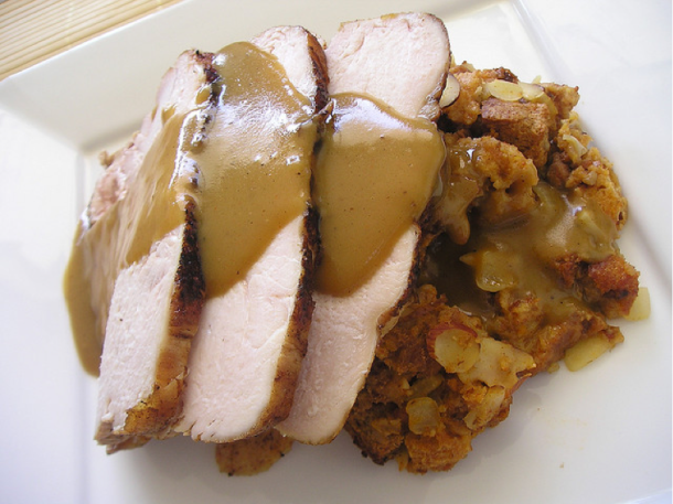
duck meat

Photo credit: Harlanh (Flickr) Duck breast – an example of dark meat

White muscle fibers, on the other hand, are specialized for more sporadic and brief energetic demands. These tissues use oxygen to burn glycogen, but can also produce energy anaerobically if needed. However, anaerobic metabolism results in the buildup of lactic acid and limits the endurance of these tissues, which is why they can only be used for short periods. White muscle fibers, unsurprisingly, are what comprise white meat—chicken breast, turkey breast, frog legs, and rabbit meat.

whitemeat

Photo credit: allthingschill (Flickr) Turkey breast

The link between structure, function, and taste can be used to answer the question of why chickens and turkeys have a combination of dark and white meat, but why ducks and geese are all dark meat. They’re all birds, after all, so it’s rather surprising that there’s such a drastic difference in their breast meat, until you consider their habits. Chickens and turkeys are relatively flightless birds. They stand, walk, or run, and use their legs to bear their weight. Their constantly used legs are mainly dark meat and their infrequently used breasts are composed of white meat. Duck and geese, however, are migratory birds whose flight patterns enlist the use of breast muscles to help them stay airborne for extended periods. To aid them in sustained flights, their chests muscles require increased stores of oxygen and myoglobin, making their breast meat dark, in stark contrast to a chicken or turkey’s breast.

The dichotomy of metabolic demands between red and white meat clearly impacts their physiology, but how does this translate into taste? The complexity of the biochemical equipment needed to store fat and oxygen and metabolize fat in red muscle tissue equates to a higher number of enzymes present in the cell that can break down and produce more flavorful compounds when cooked. A piece of dark or red meat is fattier, boasts richer and more complex flavors, and retains moisture far better than white meat.

How else can chemistry explain the different tastes of meat, ranging from the beefy flavors of cow to the gamey flavors of duck breast? It’s a common saying in the culinary world that where there’s fat, there’s flavor. While fat serves as storage for energy, it doubles as storage for flavor. With fat distributed throughout the meat, any fat-soluble flavor or aroma compounds can end up in them providing meat with its unique flavor profile. What winds up in the fat is heavily influenced by diet – cows taste “beefy” as a result of flavor compounds metabolized from grass and forage. Lamb and sheep’s distinctive flavors come from compounds produced by the liver, and the gamey flavors of duck meat are likely derived from intestinal microbes.

Examining meat through a scientific lens allows us to relate some common mantras of biology, chemistry, and cooking: structure and function go hand in hand, fat is where the flavor is, and you are (and you also taste like) what you eat. In the context of meat, physiology and flavor are intertwined and whether you’re a fan of dark or white meat, you can surely appreciate the fascinating connection between animal physiology and flavor.


References Cited:

  1. McGee, Harold. On food and cooking: the science and lore of the kitchen. New York: Simon & Schuster, 1997. Print.

Mai NguyenAbout the author: Mai Nguyen is an aspiring food scientist who received her B.S. in biochemistry from the University of Virginia. She hopes to soon escape the bench in pursuit of a more creative and fulfilling career.

Read more by Mai Nguyen


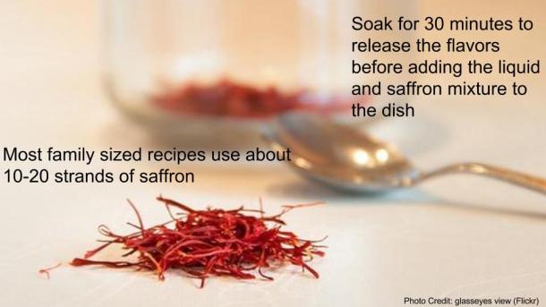
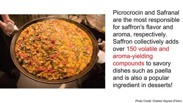
Saffron

s+f saffron post (2)

s+f saffron post

s+f saffron post (4)

s+f saffron post (5)

s+f saffron post (6)

s+f saffron post (7)

s+f saffron post (9)


Catherine HuAbout the author: Catherine Hu received her B.S. in Psychobiology at UCLA. When she is not writing about food science, she enjoys exploring the city and can often be found enduring long wait times to try new mouthwatering dishes.

Read more by Catherine Hu


Freezer Burnt Meat

Photo credit: flickr/Steven Depolo

Photo credit: flickr/Steven Depolo

Freezing is an indispensable tool in modern cooking and eating. The biochemical processes that typically occur in meats cause decay, fat oxidation, and rancidity; the higher the temperature, the faster these reactions occur. Thus, we can largely thwart off these undesirable processes by keeping meat chilled. But tossing meat into the freezer rarely results in rainbows, sunshine, or perfect burger patties, because strangely enough we can also accelerate meat decay with cold. Freezer burn can take a beautiful filet mignon and turn its surface into a leathered, unappetizing slab.

Freezer burn is caused by water sublimation from ice crystals at the meat’s surface into the dry freezer air. Sublimation occurs when a solid substance undergoes a phase change and becomes a vapor without first passing through the liquid phase. The ice crystals on the meat surface sublimate, and leave behind tiny cavities. These tiny yet numerous cavities increase the surface area of the meat and expose more tissue to the air. This accelerates oxidation of fats, which causes the rancid flavors of old spoiled meat. We usually describe oxidized fats as simply tasting “off,” which is a vague term but seems apt if you’ve ever tasted lipids past their prime, perhaps by using shortening that has been in the pantry since you were a toddler.

Photo Credit: flickr/Marcus Ward

Here, solid ice crystals directly vaporize without first passing through the liquid phase. Photo Credit: flickr/Marcus Ward

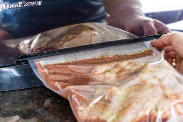
In addition to the surface area increase caused by sublimation, the freezing process itself lends itself to fat oxidation. When the liquid water in meats crystallize in the cold, the concentrations of oxidizing salts and trace metals in the tissues increases. Unfortunately, oxidation can occur over time even in wrapped and frozen meats. Some oxygen will inevitably remain in contact with the meat, unless we create a vacuum seal.

Once meat has been damaged by the cold, there’s no undoing the oxidation. So either we plan our meals so that meats are cooked immediately after purchase, or we learn to prevent the sublimation that ruins both our pork chops and our days. We simply need to keep water crystals inside the meat and keep oxygen out. Using a vacuum sealer is our best bet for avoiding freezer burn, but for cheapskates like me who won’t shell out the $30 for the sealing device, a water-impermeable plastic wrapped tightly around the meat works well enough for most home chefs.

Thus meat is sealed away happily in plastic, free from villainous oxygen. Photo credits: flickr/Mike

Thus meat is sealed away happily in plastic, free from villainous oxygen. Photo credits: flickr/Mike

References cited

  1. McGee, Harold. “Meats.” McGee on Food & Cooking: An Encyclopedia of Kitchen Science, History and Culture. London: Hodder & Stoughton, 2004. N. pag. Print.
  2. “Sublimation.” The Columbia Electronic Encyclopedia. Columbia University Press, 2012. Web. 20 July 2015.


Elsbeth SitesAbout the author: Elsbeth Sites received her B.S. in Biology at UCLA. Her addiction to the Food Network has developed into a love of learning about the science behind food.

Read more by Elsbeth Sites


Fair Food: The Science of Deep-Frying

Image Credit: (flickr/46157135@N06)

Image Credit: (46157135@N06/flickr)

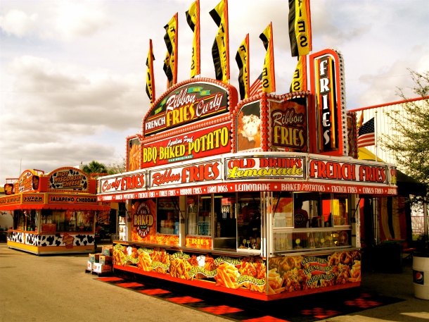
If you spent a single day at the county fair this summer, you’ll agree that the ferris wheels, petting zoos, and live music were all worth the visit. But the most exciting attraction? Fair food.

(flickr/jerkalertproductions)

Image Credit: (jerkalertproductions/flickr)

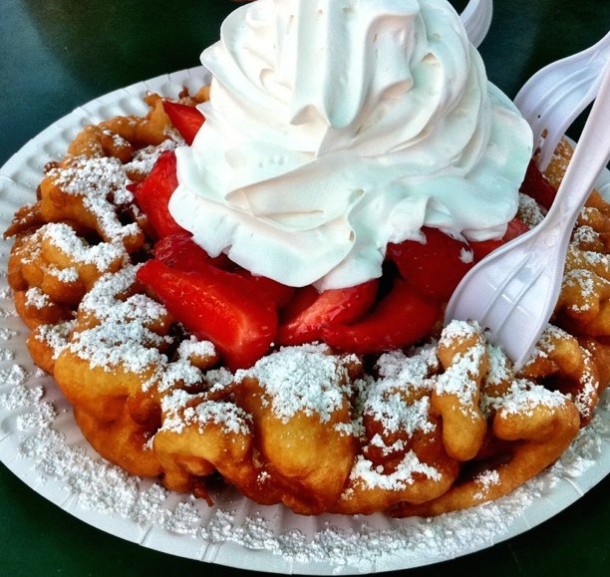
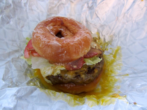
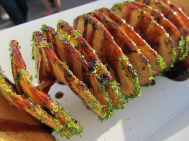
There are the classics – corn dogs, ice cream, funnel cake – but each year brings new, wacky, unbelievable, and outrageous food creations that only seem justified to consume on a hot carefree summer day at the fair. Many of this year’s jaw-dropping creations were made with the ever so popular method of deep-fat frying.

5971601817_8ce68af50d_o

Classic Funnel Cake. Image Credit: (angryjuliemonday/flickr)

Krispy Kreme Donut Cheeseburger. Image Credit: (flickr/loozrboy)

Krispy Kreme Donut Cheeseburger.
Image Credit: (loozrboy/flickr)

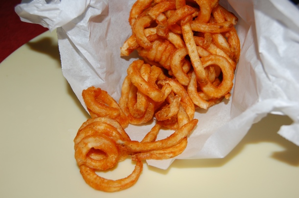
Fried Tornado Potato. Image Credit: (flickr/loozrboy)

Fried Tornado Potato.
Image Credit: (loozrboy/flickr)

Fried Moon Pie. Image Credit: (flickr/davidberkowitz)

Fried Moon Pie.
Image Credit: (davidberkowitz/flickr)

Why do deep-fried foods taste so good?
Various chemical and physical changes occur in deep-frying, including the
Maillard reactionwhich causes the aromatic browning to occur on the crunchy crust of a deep-fried treat. But first, a series of complex processes involving heat and mass transfer must occur between the food and the frying oil.

The process of deep-frying can be divided into four stages: (1) initial heating, (2) surface boiling, (3) decreasing heat transfer rate, and (4) bubble end point [1].  I will henceforth refer to the item being fried as “the food”, whether it’s a Twinkie, or Potato Chips, or Onion Rings, or Bacon-Wrapped Something On-a-Stick.

(1) Initial heating. In the first stage, the food is completely submerged into the hot oil, until the surface of the food reaches the boiling point of water. This stage lasts for about 10 seconds [1]. At this point, the heat from the oil is transferred to the food’s surface by diffusion and also by convection – a process which moves heat due to the bulk circulation of the oil’s currents from a warmer region to the cooler region surrounding the food. While convection uniformly heats the food’s surface, it doesn’t cook the center of the food. Rather, the food’s center is heated through conduction, the process by which heat diffuses from the food’s hot surface into its core through the physical contact of molecules and transfer of their thermal energy. Therefore, convection efficiently heats the food’s surface to facilitate the conduction that actually cooks the inside of the food [2].

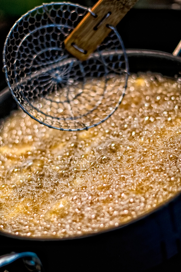
(2) Surface boiling. In this stage, we see tiny exploding bubbles sizzling at the surface of the food. Contrary to popular belief, this doesn’t mean that the oil is boiling. Instead, the hot oil surrounding the food causes water inside the food to evaporate, so the little bubbles surface as bursts of steam escaping to the food’s exterior (think jacuzzi steam jets). The movement caused by the bubbling circulates the currents of the frying oil, which increases the rate of heat transfer by “forced convection” and cooks the food faster [1].

Image Credit: (flickr/tibbygirl)

Image Credit: (tibbygirl/flickr)

These steam bubbles are important because they form a “steam barrier” around the food that repels the oil at the surface and prevents the oil from diffusing into the food, which would otherwise turn your crunchy fried treats into a soggy, greasy mess [4]. As the moisture leaves the food, the deep-fried crust we know and love begins to form!

(3) Decreasing heat transfer rate. As the crust continues to dehydrate, it conducts less heat to the rest of the food, so the rate of heat transfer through escaping steam to decreases (reduced bubbling) [1]. The remaining moisture inside of the food is slowly heated to the boiling point of water, which cooks the food inside as if it were boiled, gelatinizing the starch and denaturing the proteins in the food [3]. Now, most of the moisture from the food is lost.

(4) Bubble end point. This is the last stage of deep-frying, in which very few bubbles appear on the surface of the fried food. At this stage, water from inside the food is no longer evaporating, either because all the water from inside the food is gone, or heat transfer from the crust to the core has reduced to the point where it becomes improbable that the water will evaporate [1]. At this point, the fried product should to be removed from the oil, or else the oil will begin to seep into the fried product and make it soggy, since there are no more water vapor bubbles to counteract the diffusion of oil inwards.

Image Credit: (flickr/alexandratx)

Image Credit: (alexandratx/flickr)

Fried ice cream? Fried pizza? Fried Nutella? Armed with the science of deep-frying, the real question is, what can’t you fry?

References Cited:

  1. Farkas, B.E., Singh, R.P., Rumsey, T.R. Modeling heat and mass transfer in immersion frying. I, Model development. Journal of Food Engineering. 1996; 29(2): 211–226.
  2. Zimmerman, B. Heat Transfer and Cooking. Cooking for Engineers, [Online] June 2007.
  3. Alvis, A., Velez, C., Rada-Mendoza, M., Villamiel, M., Villada, H.S. Heat transfer coefficient during deep-fat frying. Food Control. 2009; 20: 321–325.
  4. Greene, A. Back to Basics: The Science of Frying. Decoding Delicious, [Online] May 2013.

Eunice LiuAbout the author: Eunice Liu is studying Linguistics at UCLA. She attributes her love of food science to an obsession with watching bread rise in the oven.

Read more by Eunice Liu


Cotton Candy

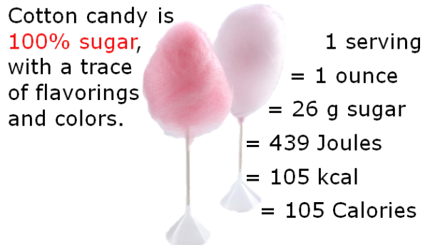
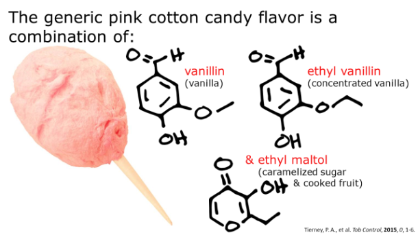
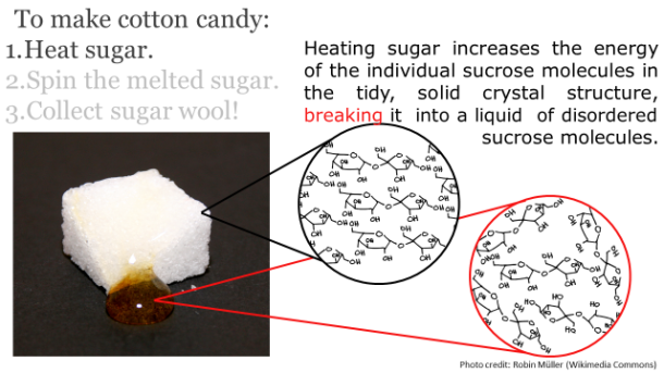
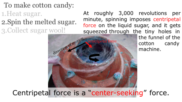
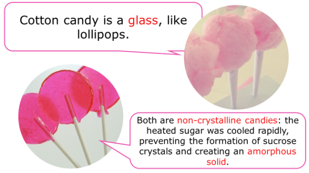
Summer would be incomplete without carnivals and bright, fleecy, sugary cotton candy. For a snack that’s nothing but sugar and air, there’s a surprising amount of physics and chemistry involved. Below are seven science-heavy facts about this feathery-light confection.

Fotm, cotton candy slide 1


Fotm, cotton candy slide 2


Fotm, cotton candy slide 3


Fotm, cotton candy slide 4


Fotm, cotton candy slide 5


Fotm, cotton candy slide 6


Fotm, cotton candy slide 7


Editor’s note: The original post stated that 1 ounce of cotton candy is 0.105 kilocalories, when in fact, it is 105 kilocalories, which is equivalent to 105 Calories. Thanks to our astute reader, Allison of the Internet for catching that! The post has now been updated (08-18-2015 10:06 p.m. PST)


Alice PhungAbout the author: Alice Phung once had her sights set on an English degree, but eventually switched over to chemistry and hasn’t looked back since.

Read more by Alice Phung


The Wonders of Baker’s Yeast

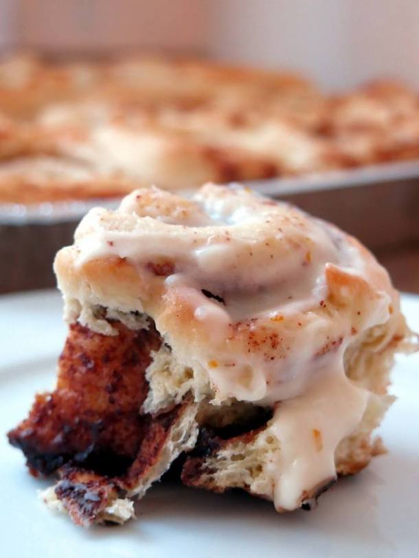
Among life’s simplest joys: smelling freshly baked cinnamon rolls wafting through the kitchen, sliding the tray of artfully coiled pastries from a warm oven, and marveling at their golden crust and fluffy interior. An ideal cinnamon roll features a potent cinnamon-sugar mixture oozing in sticky spirals. It’s often topped with a generous smear of tangy cream cheese icing that’s tempered with notes of orange peel and vanilla, sweet and rich enough to catapult you back to childhood. While the filling and icing are notable qualities, what really makes or break a cinnamon roll is its texture. Cinnamon rolls may simply serve as a vehicle for sugar and icing, but their bready foundation boasts an often-understated value. Imagine greedily lunging for a roll and biting into it, only to discover that it’s a rock-hard spiral of disappointment, instead of an airy and delicate pastry with a tender crumb. The science behind the texture of a perfectly fluffy cinnamon roll lies in the yeast.

Photo credit: Mai Nguyen

When you’re browsing the baking aisle in the grocery store, you may be overwhelmed or confused by the sheer number of different forms of yeast available—you’ll find loose granules in packets and jars, bricks, discs, and fast-rising, instant, or active dry. Despite the multitude of forms the yeasts can come in, they’re all merely purified and processed versions of the same organism. Saccharomyces cerevisiae, or baker’s yeast, is a microorganism used in professional and home kitchens alike primarily as a leavening agent for baked goods (1).

The three most commonly/commercially available forms of yeast are:

  • Caked yeast: This moist block consists of fresh, living cells that are packed tightly together. This form of yeast shows substantially higher leavening activity than its dried forms. Caked yeast is highly perishable and has a shelf life of only one to two weeks. More commonly, you’ll encounter yeast granules in packets or jars, widely available as active dry or instant.
  • Active dry: Active dry is a granular form of yeast that has been dried at high temperatures. These granules are comprised of yeast clusters that are encapsulated in a protective coating of yeast debris that formed on the surface of the granules during the drying process. These yeast cells are dormant and need to be rehydrated in warm water before being used. Simply sprinkle the granules in warm water (around 110°F), stir, and wait five to ten minutes. Water will dissolve the protective coating surrounding the granules, releasing the revived yeast cells from within. As the yeast become active, you should see a foamy layer of bubbles forming at the surface, which is carbon dioxide being released.
  • Instant rapid-rise yeast: Boasting higher viability and increased CO2 production, instant rapid-rise yeast is dried at more gentle temperatures than active dry, so more yeast cells survive this drying step. Bakers can add instant rapid-rise yeast directly to the flour, eliminating the need for prehydration. Because instant rapid-rise yeast produces carbon dioxide more vigorously than active dry yeast, these two forms of yeast should not be used interchangeably.

Granules of active dry yeast
Photo credit: Mai Nguyen

Instant rapid-rise yeast
Photo credit: Mai Nguyen

How does this tiny organism transform a dense blob of dough into a puffy masterpiece? To harness its leavening power, we rely on the phenomenon of fermentation. In the first steps of bread baking, water, yeast, flour, and salt are combined. Kneading hydrates the flour and after just a few minutes of manipulation, the dough becomes noticeably stretchier and more pliable. Water enables individual protein molecules in the dough, glutenin and gliadin, to link together to form long, elastic chains of a protein called gluten. These individual gluten strands combine to form a mesh-like network which gives bread its structure and chewy texture (2). Meanwhile, the addition of water also activates enzymes in the flour known as amylases which break down the flour’s starches into simple sugars, providing food for the yeasts (3).

The yeasts feed on these simple sugars and convert them into ethanol and carbon dioxide gas (CO2). This is where the magic begins. As carbon dioxide is released into the dough, it becomes trapped in the gluten matrix. As more and more CO2 bubbles form, the protein network stretches, inflating the dough. Depending on the recipe, dough can spend between an hour to several days rising and can expand two to four times its original size. This initial rising step is often referred to as bulk fermentation.

Like many other types of yeasted breads, a classic yeast-based recipe for cinnamon rolls calls for two rising steps. After the dough has been kneaded and has undergone bulk fermentation, it’s time to roll out the dough and shape it to prepare it for the second rising step, known as proofing. Many recipes for yeasted breads will instruct you to “punch down” dough after the initial rise. In this step, we turn and fold the dough, fill it with a cinnamon-sugar mixture, shape it into coils, and allow them to rise into bloated versions of their former selves (2). This “punching down” or turning step serves a couple of purposes: it stretches the gluten and expels excess CO2 buildup trapped in the dough from the bulk fermentation step, which can inhibit any further yeast activity. Handling the dough at this stage also redistributes yeast, moisture, heat, and sugars throughout the dough for optimal lift and flavor.

A noteworthy point: while our goal is to encourage yeast proliferation and to optimize the production of CO2 and flavor molecules, bakers should be cautious of overfermentation. If yeast fermentation happens too rapidly or continues for too long, gas bubbles can overinflate and burst, causing our dough to collapse (3). The excess of CO2 can also cause the yeast to leave behind many unwelcome tasting flavor compounds and the bread may end up tasting like alcohol.

In our final phase, our twice-risen dough is placed into the oven. Once inside, the dough experiences one last rise thanks to the high heat. The heat causes CO2 present in the dough to expand and for about the first ten minutes in the oven, the rising temperatures stimulate a rapid burst of activity in the yeast, causing them to produce even more CO2. Water and ethanol byproducts in the dough will also expand during heating. This causes the bread to rise dramatically in the oven a phenomenon known as oven spring (3). Eventually, the CO2 and alcohol are expelled from the bread and the yeast cells succumb to a dry, hot death once temperatures exceed 140°F (2).

11133670_10153200537459324_5020321292052063017_n

Photo credit: Mai Nguyen

Behind a cinnamon roll—or any kind of yeast bread —lies an intricate chemistry involved in its creation. Without the wonders of yeast and fermentation, bread wouldn’t exist as we know it today.

References Cited

  1. McGee, Harold. On food and cooking: the science and lore of the kitchen. New York: Simon & Schuster, 1997. Print.
  2. Crosby, Guy. The Science of Good Cooking. Brookline, MA: Cook’s Illustrated, 2012. Print.
  3. Bernstein, Max. “The Science of Baking Bread (And How to Do It Right).”Serious Eats. 1 Oct. 2014. Web. 11 Aug. 2015.

Mai NguyenAbout the author: Mai Nguyen is an aspiring food scientist who received her B.S. in biochemistry from the University of Virginia. She hopes to soon escape the bench in pursuit of a more creative and fulfilling career.

Read more by Mai Nguyen


Jammin’ With Fruit

Now that summer is in full swing, what better way is there to use all the berries, apricots, plums, peaches, and nectarines in season than to make jam? Jam has a great texture that makes it the perfect spread for brioche toast and a sweet complement for porridge. The base ingredients of fruit, sugar, pectin, and acid are cooked until the jam reaches a spreadable consistency [1]. Each ingredient plays an important role in the texture of the final product.

Strawberry jam. Photocredit: Julia Khusainova (Jullclous/Flickr)

Strawberry jam. Photocredit: Julia Khusainova (Jullclous/Flickr)

Sugar Skills

Sugar has many roles besides adding sweetness to the jam. When sugar is mixed with mashed fruit, it begins to dissolve and draw water out of the fruits through osmosis [1]. This occurs because fruit has a lower concentration of sugars than the amount of sugar that is typically added. The hydrophilic groups on sugar make it miscible with other polar molecules like water. Sugar also acts as a preservative by forming bonds with water molecules, making fewer water molecules available to support the growth of various microorganisms that might cause spoilage, such as Aspergillus glaucus and Saccharomyces rouxii [2,3].

5229543600_661472e1e3_o

The making of blackberry jam. Photocredit: (mrskupe/Flickr)

Gelation

Pectin is soluble dietary fiber that naturally occurs in certain fruits such as apples, plums, and quinces. When heated and mixed with acid, this carbohydrate creates a thick gel that contributes to the consistency of jam. Acid from citrus increases the hydrogen ion concentration in the solution, which results in the pectin molecules losing some charge. With less electrostatic repulsion, the molecules can now aggregate to form a physical gel at a higher temperature of around 220ºF [4], resulting in watery fruit liquid dispersing itself within a web of pectin molecules [5].

Typically, under-ripe fruits have more pectin because fruit enzymes convert it to pectic acid during the ripening process [5]. This means that high pectin content can often be a trade-off for lower flavor, so it is recommended to use two parts of ripe fruit for every part of under-ripe fruit for the best consistency and taste [1]. Alternatively you can buy powdered pectin from the store to use with fruits that naturally have low levels of this carbohydrate, such as apricots, peaches, and raspberries.

Sugar Inversion

Acid not only contributes to the texture of jam, but it also catalyzes the conversion of sucrose (from added sugar) into its constituent fructose and glucose molecules with the help of heat. This process is called sugar inversion, and it is necessary to prevent recrystallization during jam storage [6]. However, this rarely occurs because finishing a jar of jam usually does not take too long. As an added bonus, acid also contributes to the flavor balance of the jam, preventing it from being too sweet.

Different fruits vary in acid and pectin content, so adjustments may be necessary to obtain the right texture and taste. You can also use a Brix test to measure the endogenous levels of sugars and dissolved nutrients. Basically if your jam contains riper and more nutrient dense fruits, the test will give a higher reading. Develop the right recipe, and you will want to eat out of the jam jar with a spoon!

References cited:

  1. The Science of Jam and Jelly Making. University of Kentucky.
  2. Jam Making: Why all the sugar? Iufost.org
  3. Why does jam go mouldy, even in the fridge? University of Liverpool
  4. Fishman, M.L. & Jen, J.J. Chemistry and Function of Pectins. June 1986.
  5. Jam Making 101. Seriouseats.
  6. Inversion of Sucrose. Colby College.


Catherine HuAbout the author: Catherine Hu is pursuing her B.S. in Psychobiology at UCLA. When she is not writing about food science, she enjoys exploring the city and can often be found enduring long wait times to try new mouthwatering dishes.

Read more by Catherine Hu