Tag Archive for: physiology

Sanshool Seduction: The Science of Spiciness

One of the most aggressive flavors we can experience is spiciness. Imagine a bright red chili pepper whose color gives us fair warning of its propensity to ignite a fire. In fact, a common physiological response to eating spicy food is analogous to the way our body responds to an elevation in internal body temperature. You can feel the burn. The consumption of spicy ingredients triggers our exocrine (sweat) glands to secrete fluid at the skin’s surface to promote cooling through the evaporation of our perspiration.

So how is it that spicy food can make us sweat?

capsician

Red Chili Peppers [Photo Credit: The Paleo Diet]

capsaicin2

Chemical Structure of Capsaicin [Photo Credit: Dartmouth]


The answer lies in our head.  Our body’s reaction to spiciness is a result of input delivered from receptors in our mouth to our central nervous system. A compound found in many spicy ingredients such as chili peppers is called capsaicin.  This molecule activates sensory neurons in our mouth called thermal nociceptors [1,2]. The activation of these thermal pain receptors in turn stimulates our sympathetic nervous system [3], which is associated with our body’s “flight-or-flight” responses.  Therefore, the characteristic increase in heart rate and sweat production due to the consumption of capsaicin-rich ingredients is due in part to the capsaicin’s role in adrenaline secretion [4].

If you decide to brave the spice, here’s some advice: When it comes to relieving the pain of the capsaicin burn, your best bet is to have a glass of cold milk or better yet, a bowl of ice cream.  The compound, casein, is a hydrophobic substance found in milk that operates to “absorb” the lipid-rich capsaicin molecules making it easier to cleanse your mouth of the spicy chemical [5].

However, capsaicin isn’t the only fiery compound that can elicit unique physiological responses. On the other end of the spectrum is a compound known as hydroxyl-alpha sanshool, commonly found in Z. Bungaenum peppercorns.  This variety of peppercorn is commonly grown in the Sichuan province of China, and is referred to as the Sichuan peppercorn.  The characteristic “flavor sensation” one might experience when eating Sichuan cuisine is a mind-numbing spiciness.

Here’s how that happens.

Sichuan Peppercorns [Photo Credit: Serious Eats]

Sichuan Peppercorns [Photo Credit: Serious Eats]

Chemical Structure of Hydroxy-Alpha Sanshool [Photo Credit: Hong Kong University]

Chemical Structure of Hydroxy-Alpha Sanshool [Photo Credit: Hong Kong University]

Hydro-alpha sanshool activates somatosensory neurons that are responsible for detecting innocuous stimuli such as a gentle touch. This is opposite of the nociceptors that detect capsaicin which primarily detect noxious or painful stimuli.  The stimulation of somatosensory neurons by hydro-alpha sanshool produces a similar effect to local anesthetics used to moderate pain in surgery.   The compound found in Sichuan peppercorns trigger somatosensory neurons to prevent the influx and efflux of electrolytes, Na+ and K+, through ion channels [6,7].  The resulting effect of hydro-alpha sanshool is to cease the propagation of neuronal action potential through the nervous system’s pain pathways.  So, when you enjoy a delicious dish of Liang Fen (Cold Mung Bean Noodles with Sichuan Peppercorn/Chili Vinegar), or Suan Cai Yu (Poached Fish in Green Sichuan Peppercorn Sauce) you can experience thrilling mouth numbness. Your first instinct after eating these dishes will be to grab the closest glass of water available to you.  Interestingly, a sip of flat water after eating these dishes will produce a surprising fizziness like you might experience while drinking sparkling water.

Liang Fen: Cold Mung Bean Noodles with Sichuan Peppercorn/Chili Vinegar [Photo Credit: Serious Eats]

Liang Fen: Cold Mung Bean Noodles with Sichuan Peppercorn/Chili Vinegar [Photo Credit: Serious Eats]

Liang Fen: Cold Mung Bean Noodles with Sichuan Peppercorn/Chili Vinegar [Photo Credit: Serious Eats]

Liang Fen: Cold Mung Bean Noodles with Sichuan Peppercorn/Chili Vinegar [Photo Credit: Serious Eats]

References cited

  1. Caterina, M., Schumacher, M., Tominaga, M., Rosen, T., Levine, J., Julius, D. “The capsaicin receptor: a heat-activated ion channel in the pain pathway”. Nature. 389 (1997): 816-824.
  2. Liu, M., Max, M., Parada, S., Rowan, J., Bennett, G. “The Sympathetic Nervous System Contributes to Capsaicin-Evoked Mechanical Allodynia But Not Pinprick Hyperalgesia in Humans” The Journal of Neuroscience. 16.22 (1996): 7331-7335.
  3. Ohnuki, K., Moritani, T., Ishihara, K., Fushiki, T. “Capsaicin Increases Modulation of Sympathetic Nerve Activity in Rats: Measurement Using Power Spectral Analysis of Heart Rate Fluctuations”. Bioscience, Biotechnology, and Biochemistry. 65.3 (2001): 638-643.
  4. Watanabe, T., Sakurada, N., Kobata, K. “Capsaicin-, Resiniferatoxin-, and Olvanil-Induced Adrenaline Secretions in Rats Via the Vanilloid Receptor” Bioscience, Biotechnology, and Biochemistry. 65.11 (2001): 2443-2447.
  5. Fire and Spice. General Chemistry Online!.
  6. Tsunozaki, M., Lennertz, RC., Vilceanu, D., Katta, S., Stucky, CL., Bautista, DM. “A ‘Toothache Tree’ Alkylamide Inhibits AD Mechanonociceptors to Alleviate Mechanical Pain” Journal of Physiology. 591 (2013) 3325-3340.
  7. Bautista, DM., Sigal, YM., Milstein, AD., Garrison, JL., Zorn, JA., Tsuruda, PR., Nicoll, RA., Julius, D. “Pungent Agents from Szechuan Peppers Excite Sensory Neurons by Inhibiting Two-Pore Potassium Channels” Nature Neuroscience. 11.7 (2011): 772-779.

Anthony MartinAbout the author: Anthony Martin received his Ph.D. in Genetic, Cellular and Molecular Biology at USC and is self-publishing a cookbook of his favorite Filipino dishes.

Read more by Anthony Martin


Meat: where physiology meets flavor

A charcuterie board is the perfect accompaniment to any gathering and rivals a cheese plate as a crowd-pleaser. It’s low maintenance, delicious, and will almost certainly have a taste or texture to appeal to the pickiest of palates. Meat comes in an array of textures, fat content, and flavors, which vary species to species and even within the same animal. Flavor profiles of meat can vary wildly and subtleties between different cuts of meat can all be largely explained by chemistry.

meat

Photo credit: willmacdonald18 (Flickr)

What is meat exactly? Meat can loosely mean any type of edible tissue originating from an animal – including everything from chicken feet to cow tongue. The majority of meat we consume, however, is skeletal muscle tissue, comprising roughly 75% water, 20% protein, and 3% fat. While the function of muscle tissue—which is to generate movement—is simple, muscle tissue is a complex system of biochemical machinery.

Muscle tissue consists white and red fibers, which each generate contrasting types of movement. The major differences between the two types of muscle fibers are summarized in the table below.

red muscle fibers white muscle fibers
alternate names slow-twitch fibers fast-twitch fibers
relative size thin thick
movement type prolonged, deliberate short, high-powered bursts
fueled by fat + oxygen glycogen
Requires oxygen? absolutely yes, but can run anaerobically
taste/nutrtion fattier, more flavorful higher in protein, relatively bland
appearance dark/red white

The main distinction between these types of tissue is in their function and metabolic demands. Red muscle fibers are designed for endurance—think long distance running—or sustained motion. Red tissue runs on fat and oxygen and absolutely requires oxygen to function properly. Since red muscle tissue demands oxygen, it contains an abundance of a pigmented protein called myoglobin. Myoglobin binds and stores oxygen from the then passes it along to a fat-oxidizing cytochrome that generates ATP to fuel the cell. The more exercise a muscle receives, the higher its demand for oxygen, and the more myoglobin and cytochromes it will contain, leading to a darker appearance.

duck meat

Photo credit: Harlanh (Flickr) Duck breast – an example of dark meat

White muscle fibers, on the other hand, are specialized for more sporadic and brief energetic demands. These tissues use oxygen to burn glycogen, but can also produce energy anaerobically if needed. However, anaerobic metabolism results in the buildup of lactic acid and limits the endurance of these tissues, which is why they can only be used for short periods. White muscle fibers, unsurprisingly, are what comprise white meat—chicken breast, turkey breast, frog legs, and rabbit meat.

whitemeat

Photo credit: allthingschill (Flickr) Turkey breast

The link between structure, function, and taste can be used to answer the question of why chickens and turkeys have a combination of dark and white meat, but why ducks and geese are all dark meat. They’re all birds, after all, so it’s rather surprising that there’s such a drastic difference in their breast meat, until you consider their habits. Chickens and turkeys are relatively flightless birds. They stand, walk, or run, and use their legs to bear their weight. Their constantly used legs are mainly dark meat and their infrequently used breasts are composed of white meat. Duck and geese, however, are migratory birds whose flight patterns enlist the use of breast muscles to help them stay airborne for extended periods. To aid them in sustained flights, their chests muscles require increased stores of oxygen and myoglobin, making their breast meat dark, in stark contrast to a chicken or turkey’s breast.

The dichotomy of metabolic demands between red and white meat clearly impacts their physiology, but how does this translate into taste? The complexity of the biochemical equipment needed to store fat and oxygen and metabolize fat in red muscle tissue equates to a higher number of enzymes present in the cell that can break down and produce more flavorful compounds when cooked. A piece of dark or red meat is fattier, boasts richer and more complex flavors, and retains moisture far better than white meat.

How else can chemistry explain the different tastes of meat, ranging from the beefy flavors of cow to the gamey flavors of duck breast? It’s a common saying in the culinary world that where there’s fat, there’s flavor. While fat serves as storage for energy, it doubles as storage for flavor. With fat distributed throughout the meat, any fat-soluble flavor or aroma compounds can end up in them providing meat with its unique flavor profile. What winds up in the fat is heavily influenced by diet – cows taste “beefy” as a result of flavor compounds metabolized from grass and forage. Lamb and sheep’s distinctive flavors come from compounds produced by the liver, and the gamey flavors of duck meat are likely derived from intestinal microbes.

Examining meat through a scientific lens allows us to relate some common mantras of biology, chemistry, and cooking: structure and function go hand in hand, fat is where the flavor is, and you are (and you also taste like) what you eat. In the context of meat, physiology and flavor are intertwined and whether you’re a fan of dark or white meat, you can surely appreciate the fascinating connection between animal physiology and flavor.


References Cited:

  1. McGee, Harold. On food and cooking: the science and lore of the kitchen. New York: Simon & Schuster, 1997. Print.

Mai NguyenAbout the author: Mai Nguyen is an aspiring food scientist who received her B.S. in biochemistry from the University of Virginia. She hopes to soon escape the bench in pursuit of a more creative and fulfilling career.

Read more by Mai Nguyen


Physiology of Foie Gras

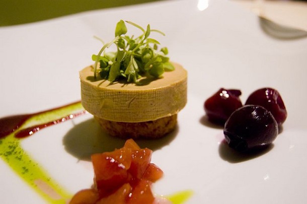
Photo Credits: (flickr/Ulterior Epiculture)

Photo Credits: (flickr/Ulterior Epicure)

Decadent, diseased, silky, sinful. The adjectives that follow foie gras range from the disgusting to the luxurious. The fattened liver of a duck or goose polarizes people, and there seems to be no middle ground wherein a person can both enjoy foie gras and ethically question it. Because it is such a controversial food, the discourse surrounding it is often steeped in emotion, but the best way to make an informed, fact-based decision is through science. Here we will examine physiology, pathology, and a bit of genetics regarding waterfowl and foie gras in an attempt to promote overall awareness of what we eat (or don’t eat).

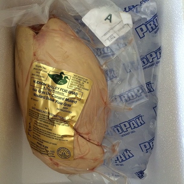
Foie gras is French for fatty liver, and that is exactly what it is. The liver of a bird, usually a duck or sometimes a goose, that has been force-fed to the point of having a fat, enlarged liver. The liver must weigh more than 300g for ducks, and 400g for geese to legally bear the name foie gras in France. [1] Force-feeding is typically done through a practice called gavage, wherein a long tube is inserted into the bird’s mouth and throat up to three times a day for 3-10 days. French rural code L654-27-1 states that “Foie gras belongs to the protected cultural and gastronomical heritage of France.”[2] Currently, the farming of animals to produce foie gras is banned in 22 EU nations, but not its sale or import. In California, the sale of foie gras was banned in 2004, the ban was lifted in early 2015 by a federal court, and the lifting of that ban is currently being appealed. [3] Needless to say, it’s a very complicated issue.

Photo Credits: (flickr/Pickled newt)

Photo Credits: (flickr/Pickled newt)

Foie Gras Physiology

Many bird species, including ducks and geese, eat prey much wider than the diameter of their esophagus. Consequently, the inner diameter of the upper part of the esophagus is comparatively larger than in mammals. [4] Also unlike mammals, the upper esophagus is not circled by cartilaginous rings, which explains how birds swallow whole, live fish with ease. In humans, the upper esophageal sphincter is a high-pressure zone situated between the pharynx and the cervical esophagus. The sphincter is composed of muscle, cartilage, and bone, [5] and thus is much more rigid than the upper esophagus of a waterfowl. Most birds species posses an “outpouching” of the esophagus, known as the crop. It also allows the birds to store large amounts of food before sending it along to the stomach for digestion. [4]

Another important difference in human and duck anatomy is the trachea. In humans, food and air start along the same path in the mouth, then the trachea (or windpipe) branches off at the back of the throat where the epiglottis prevents food from entering the trachea and channels swallowed food along its proper route, the esophagus. Try to force something past the epiglottis, and you trigger the unpleasant pharyngeal reflex, or gag reflex. Certainly a gavage would trigger this in a human, which is one reason why images of force fed birds make us so uncomfortable. Foie producers say if the procedure is carried under proper conditions, the gavage does not block the upper respiratory tract as the birds’ tracheas and esophaguses are completely separate, [6] and thus they do not gag or feel discomfort as a human would. However, foie gras critics rebuke that this is a ridiculous excuse, and that the birds are clearly harmed by the gavage.

Ducks and geese are sometimes reported to be panting when observed in a foie-farm. But before we assume they do so because they’re in distress, we should keep in mind that like a dog, panting in birds is a thermo-regulatory reflex. [4] Humans have sudoriferous glands (sweat glands) that discharge sweat to take care of latent heat, but birds do not. They regulate body temperature by opening their beaks and panting to cool down. Researchers have examined whether other avian behaviors are indicators of distress, like avoidance behavior, elevated heart rate, or elevated cortisol (stress hormone) levels. They report that force-feeding does not stress the birds more than typical capture and handling does. [1] [Side note: Most of this research was conducted by the same group of scientists from the French National Institute for Agricultural Research, so it would be helpful to have experiments performed by more organizations.]

Photo Credits: (flickr/Jeremy Couture)

Photo Credits: (flickr/Jeremy Couture)

Pathology

In mammals, hepatic steatosis (fatty liver) is a pathological condition. Human fatty liver occurs when there is an imbalance of fat uptake and export in the liver, most often caused by alcoholism, malnutrition, obesity, or diabetes. On its own, hepatic steatosis is not harmful and can be reversed, but if not addressed with dietary and lifestyle change it can develop into cirrhosis, wherein the healthy liver tissue is replaced by scar tissue, or necrosis (tissue death). Indeed, foie gras in a human is a disease. [7]

Certain metabolic adaptations in migratory birds and fish cause a natural hepatic steatosis, and proponents of foie gras use this observation to argue that the condition is not pathological in those species. These animals must compile large energy stores for their migrations, and they do so by ingesting carbohydrates and storing the energy as fat, a process called lipogenesis. [8] Foie producers posit that they are simply exploiting the incredible lipogenetic abilities of the fowl liver. The human liver does no more than 30% of our entire bodies’ lipogenesis, as our adipose tissue carries most of the workload. [9] By contrast, the avian liver performs the vast majority of their lipogenesis, up to 96% of it in some species. [10] To further their argument that their birds are not diseased, foie farmers assert that it is in their best interest to avoid producing diseased livers, as they are of no commercial value.

Photo Credits: (flickr/Jay Tong)

Photo Credits: (flickr/Jay Tong)

Genetics of the Muscovy Duck

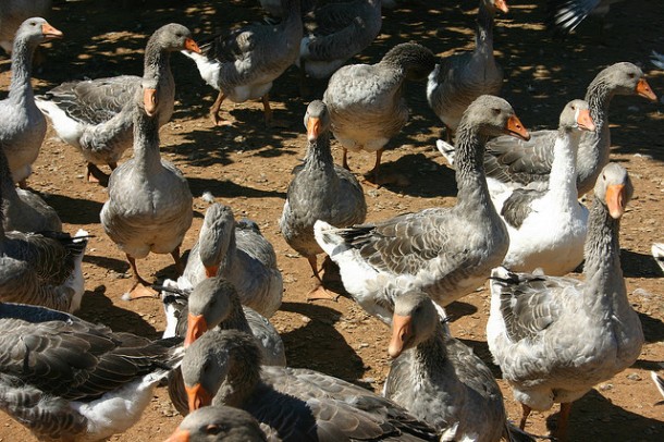
In addition to the anatomical and physiological aspects of waterfowl that may make the production of foie gras seem less cruel, a look into the breed may provide further insight. Foie gras is made from the liver of the Moulard duck, which is the product of a female Pekin artificially inseminated with the sperm of a male Muscovy duck. [1]The Moulard, or “mule duck” genotype is not present in the wild, and like other hybrid species, it is sterile. Therefore, the animals themselves cannot breed more baby Moulards.

Muscovies are non-migratory, [11] so unlike migratory species, in natural settings they do not gorge themselves to put on extra fat to carry them through long periods of physical exertion with no breaks to replenish energy. It might seem like they would be a poor choice for duck farming, but they are prized for their well-flavored, lean meat.

Pekin ducks on the other hand have many of the characteristics of migratory species. They are naturally gregarious and clump themselves together whether or not they have space to roam. [11] Years of breeding have made them very plump and small-winged, and thus they no longer migrate. However, their inner organs and basic metabolism still maintain characteristics of migratory waterfowl. The moulard thus exhibits the more desirable behavioral features of the two species. Like muscovies, they have no migratory instincts, so they are easy to farm-raise. But they retain all of the anatomy and metabolism of Pekins that naturally make them want to gorge and store energy as fat.

Photo Credits: (flickr/Taylor149)

Photo Credits: (flickr/Taylor149)

In this physiological context, gavage and foie gras might not be as tortuous as some imagine it to be. Even with this information, some people may still feel uncomfortable with the idea of force-feeding, and that is perfectly reasonable. If we want to eat foie gras entirely guilt-free, perhaps we should support the production of “humane” foie gras, where the animal is left to gorge on its own as if it were preparing for migration. Examining foie gras through a scientific lens teaches us to evaluate the animal body for its natural capabilities, but science does not always give us clear answers as to what is morally right. Regarding foie gras, the jury is literally still out.

References Cited

  1. Guémené, Daniel, Gérard Guy, Jérôme Noirault, Nicolas Destombes, and Jean-Michel Faure. “Rearing Conditions during the Force-feeding Period in Male Mule Ducks and Their Impact upon Stress and Welfare.” Animal Research 55.5 (2006): 443-58. Web.
  2. “Legifrance – Le Service Public De L’accès Au Droit.” Code Rural Et De La Pêche Maritime. N.p., n.d. Web. 19 Feb. 2015
  3. McClurg, Lesley. “The Legal Battle Over Foie Gras Continues.” – Capradio.org. Capital Public Radio, 9 Feb. 2015. Web. 19 Feb. 2015.
  4. Guémené, Daniel, Gérard Guy, Jacques Servière, and Jean-Michel Faure. “Force Feeding: An Examination of Available Scientific Evidence.” Artisan Farmers Alliance (n.d.): n. pag. Artisanfarmers.org. Web.
  5. Kuo, Braden, and Daniela Urma. “Esophagus – Anatomy and Development.” GI Motility Online (2006): n. pag. Web.
  6. http://onlinelibrary.wiley.com/store/10.1111/j.1740-8261.1991.tb00087.x/asset/j.1740-8261.1991.tb00087.x.pdf?v=1&t=i5inm4hl&s=a129cb6b04b350dfd5778a83eaaea1f2f0fb02a0
  7. Jaeschke, Hartmut, Jaspreet S. Gujral, and Mary Lynn Bajt. “Apoptosis and Necrosis in Liver Disease.” Liver International 24.2 (2004): 85-89. Web.
  8. Pilo, B., and J.c. George. “Diurnal and Seasonal Variation in Liver Glycogen and Fat in Relation to Metabolic Status of Liver and M. Pectoralis in the Migratory Starling, Sturnus Roseus, Wintering in India.” Comparative Biochemistry and Physiology Part A: Physiology 74.3 (1983): 601-04. Web.
  9. Timlin, Maureen T., and Elizabeth J. Parks. “Temporal Patterns of De Novo Lipogenesis in the Postprandial State in Healthy Men.” The American Journal of Clinical Nutrition 18.1 (2005): 35-42. Web.
  10. Desmeth, M., M. Messeyne, G. Schuermans, J. Vandeputte-Poma, and F. Vandergeynst. “Effect of Age and Diet on the Fatty Acid Composition of Triglycéridesand Phospholipids from Liver, Adipose Tissue and Crop of the Pigeon.” The Journal of Nutrition 111 (1980): n. pag. Web.
  11. Lopez, Kenji. “The Physiology of Foie: Why Foie Gras Is Not Unethical.” Serious Eats. N.p., 16 Dec. 2007. Web. 18 Feb. 2015.

Elsbeth SitesAbout the author: Elsbeth Sites is pursuing her B.S. in Biology at UCLA. Her addiction to the Food Network has developed into a love of learning about the science behind food.

Read more by Elsbeth Sites


Thanksgiving, Turkeys, and Tryptophan

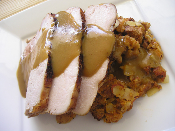
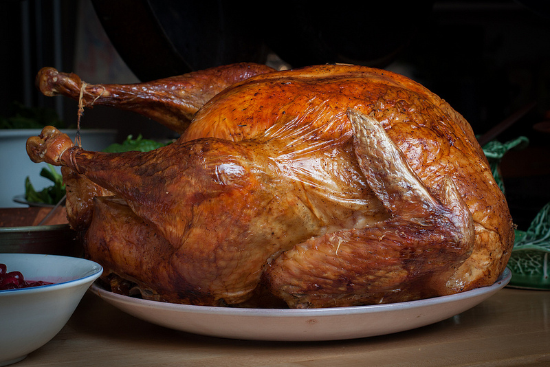
Photo credit: Tim Sackton (timsackton/Flickr)

Photo credit: Tim Sackton (timsackton/Flickr)

Turkey is the star of the most famous dinner of the year; it is also the victim of a myth that persists every holiday season. At the end of Thanksgiving dinner, there’s a good chance that someone will mention that a molecule called tryptophan is the culprit for the post-feast drowsiness. The science seems sound enough. Turkey contains tryptophan, which is a precursor for serotonin, a neurotransmitter. In turn, serotonin produces melatonin, a hormone that helps regulate sleep. This myth perpetuates, like many others, because it is based on a huge oversimplification of the truth.

On the most fundamental level, tryptophan is an essential amino acid required to make many different proteins in the body. Our bodies can’t produce tryptophan, so we have to get it from the foods we eat. Considering amino acids are used to make proteins, we get them by consuming other proteins such as meats, poultry, eggs, dairy, rice, and beans.

Chemically, tryptophan is the same whether it’s in a test tube or in our bloodstream, meaning there’s really nothing special about the tryptophan found in turkey versus other protein sources, like chicken. Turkey actually contains less tryptophan per gram than chicken, and half as much as in soybeans [1], but would anyone ever blame a tryptophan-induced food coma on a Thanksgiving chicken or tofurkey?

Tryptophan can directly cause drowsiness—if taken in pill form on an empty stomach [2]. Before tryptophan can be converted to serotonin, it must first cross the blood-brain barrier to enter the brain. This would be simple enough if tryptophan was the only amino acid capable of crossing the blood-brain barrier. However, after a meal―especially a high-protein meal like a turkey dinner―there will be many different amino acids floating around the bloodstream, many of which can also enter the brain. Despite this barrage at the gates, the brain can only take up a limited amount of amino acids. As tryptophan makes up only a small fraction of all the amino acids we consume, the other amino acids directly compete with tryptophan to cross the blood-brain barrier, ultimately decreasing tryptophan’s chances of entering the brain. Even if you stuffed yourself on nothing but tofurkey, only small amounts of tryptophan would ever enter the brain to make you sleepy.

The culprits behind the Thanksgiving dinner food coma are thus largely the high-carbohydrate dishes (and alcoholic beverages) surrounding the turkey: potatoes, yams, pies, bread, and stuffing. Eating carbohydrate-rich meals stimulates the production and release of insulin into the bloodstream [3]. Insulin then signals the uptake of amino acids into the muscles. Unlike most amino acids, tryptophan has a rather large and bulky structure that prevents it from entering muscles, so it is left behind in the bloodstream. With fewer of the other amino acids in the blood, tryptophan has less competition and is more likely to cross the blood-brain barrier, where it can be converted into serotonin and melatonin, the brain chemicals attributed to happiness and sleepiness [4].

At this point you might be thinking, “So turkey does make you sleepy! It just needs help from carbs!” Yes, but on that level, Thanksgiving dinner wouldn’t be any different from, say, a bacon and biscuits binge, or any other high-protein, high-carb meal.

And some of you might still adamantly insist, “I even eat turkey outside of Thanksgiving, and I still get super sleepy! It must be the turkey!” But remember: the sugar pill works for a reason. Never underestimate the power of the mind.

Still, don’t let any of this stop you from enjoying this holiday and stuffing your face to your heart’s content.


References Cited

  1. Foods Highest in Tryptophan. Nutrition Data.
  2. Hartmann E (1982). “Effects of L-tryptophan on sleepiness and on sleep”. Journal of Psychiatric Research 17 (2): 107–13.
  3. Wurtman RJ, Wurtman JJ, Regan MM, McDermott JM, Tsay RH, Breu JJ (2003). “Effects of normal meals rich in carbohydrates or proteins on plasma tryptophan and tyrosine ratios”. Am. J. Clin. Nutr. 77 (1): 128–32.
  4. Lyons PM, Truswell AS (1988). “Serotonin precursor influenced by type of carbohydrate meal in healthy adults”. Am. J. Clin. Nutr. 47 (3): 433–9.

Alice PhungAbout the author: Alice Phung once had her sights set on an English degree, but eventually switched over to chemistry and hasn’t looked back since.

Read more by Alice Phung